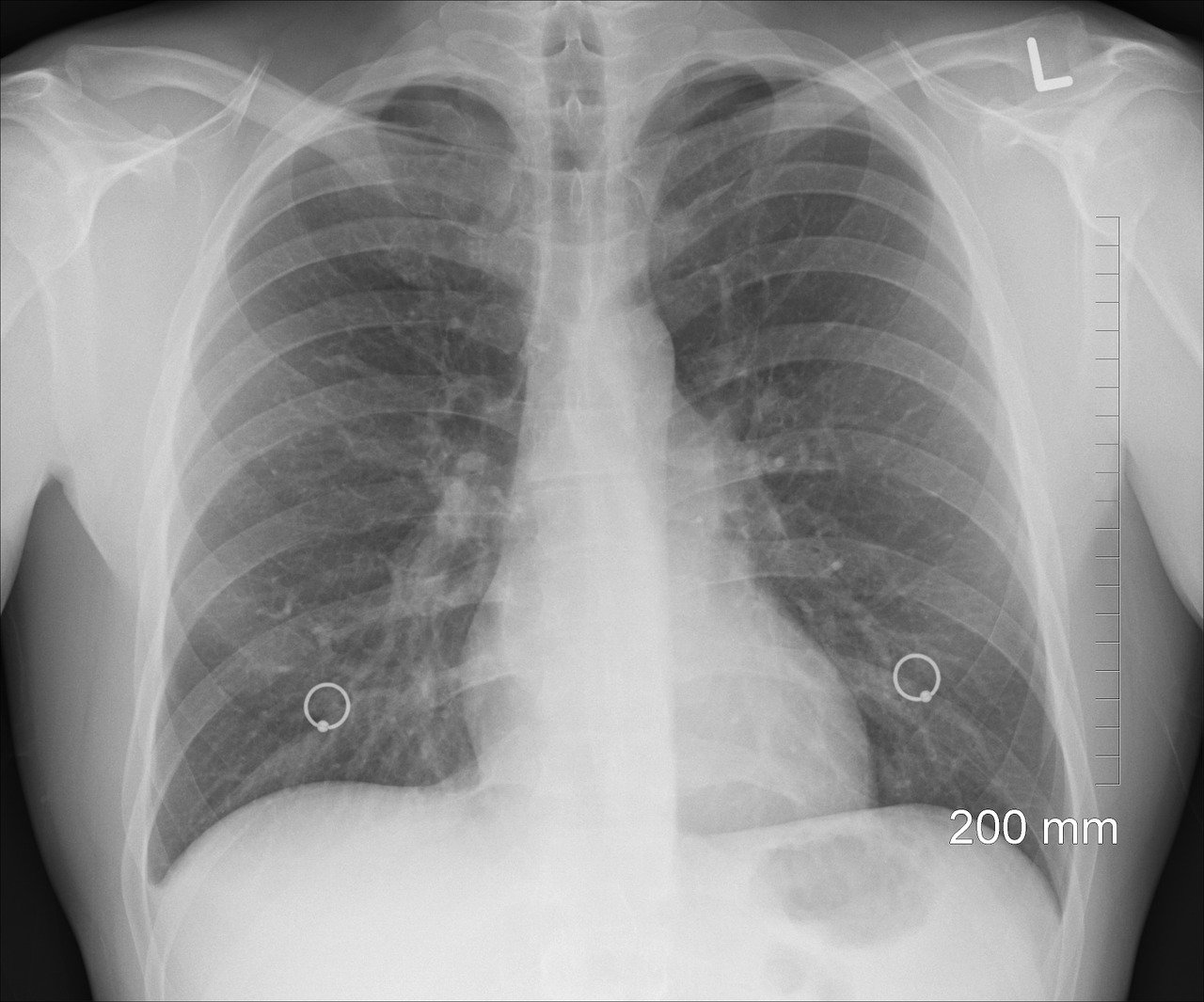
Severe cases of COVID-19 can cause pneumonia, a process whereby the delicate air sacs in the lungs swell with fluid, and their ability to take in oxygen from the air becomes dangerously compromised. In these cases, ventilators are often required to support the patient’s breathing. A full recovery from COVID-19 does not always spell the end of respiratory issues, with many experiencing long-term effects after pneumonia that take months to resolve, if at all.
Radiologists and physicians at UC San Diego Health have responded to the crisis with a diagnostic tool for predicting COVID-19 pneumonia much earlier, using an algorithm powered by artificial intelligence (AI).
This AI capability is a game-changer when it comes to rapidly and accurately predicting the health outcomes of those who have tested positive for the coronavirus. Critically, the platform not only picks up signs of full-blown pneumonia but also highlights early physiological changes in the lungs occurring in those who have yet to be diagnosed.
One particular clinical case highlighted the implications of this new platform perfectly. An individual with no symptoms of COVID-19 went for a chest X-ray for unrelated reasons. The AI tool alerted to the early signs of pneumonia and after confirmation by a radiologist, the patient was found to be positive for COVID-19 and early intervention was initiated.
The machine learning algorithm was developed by Albert Hsiao, MD, Ph.D., associate professor of radiology at the University of California San Diego School of Medicine and “taught” by a series of over 22,000 notations on chest X-ray images by human radiologists.
“Pneumonia can be subtle, especially if it’s not your average bacterial pneumonia, and if we could identify those patients early before you can even detect it with a stethoscope, we might be better positioned to treat those at highest risk for severe disease and death,” said Hsiao.
In a study published in the Journal of Thoracic Imaging, Hsiao and the team demonstrated the potential of AI at accelerating the diagnosis of COVID-related pneumonia. Published X-rays from COVID-19 patients from both China and the United States were fed into Hsiao’s AI-driven system. The algorithm hit a home run every time, consistently and correctly spotting localized areas of pneumonia in the chest X-ray images. This was especially impressive, considering the images were highly varied in terms of capture technique, image quality, contrast, and resolution. According to Hsiao, this type of technology could potentially replace existing diagnostic assays for COVID-19, identifying individuals even at the early stages of infection.
Sources: Journal of Thoracic Imaging, UC San Diego Health.
