Santa Cruz Biotechnology is a world leader in the development of products for the biomedical research market. Over the past 30+ years, the Company has focused on the ongoing development of research antibodies, siRNA and CRISPR Gene editing tools, biochemicals, labware and more recently has expanded into animal health care products. Santa Cruz Biotechnology has the highest commitment to quality and customer service.
Santa Cruz Biotechnology
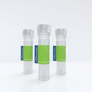
NFATc1 (7A6) Alexa Fluor® 647 | Santa Cruz Biotechnology
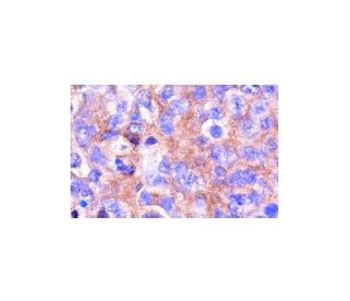
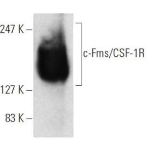
mouse monoclonal IgG1; NFATc1 Antibody (7A6) is an IgG1 κ mouse monoclonal NFATc1 antibody (also designated NFAT2 antibody, or nuclear factor of activated T cells 1 antibody) that detects the NFATc1 protein of mouse, rat and human origin by WB, IP, IF, IHC(P) and FCM. NFATc1 Antibody (7A6) is available as both the non-conjugated anti-NFATc1 antibody form, as well as multiple conjugated forms of anti-NFATc1 antibody, including agarose, HRP, PE, FITC and multiple Alexa Fluor® conjugates. Members of the NFAT (nuclear factor of activated T cells) family of transcription factors are related to NFκB/Rel proteins and form cooperative complexes with the AP-1 proteins, Fos and Jun, on DNA to regulate cytokine expression in T cells. NFAT proteins are widely expressed and alternatively modified to generate splice variants, and they are localized to both the cytosol (NFATc) and to the nucleus (NFATn). NFATc1 (NFATc), NFATc2 (NFATp) and NFATc3 (NFAT4, NFSTx) are predominantly expressed in immune cells, and NFAT2 and NFATc4 are expressed at high levels in cardiac tissues. In addition to activating cytokine gene transcription, NFATc2 is also implicated in cardiac valve development, and N FATc4 is involved in cardiac hypertrophy. NFAT5 is detected in both immune and nonimmune cells and, like other NFAT proteins, it contains a highly conserved Rel-like binding domain that mediates NFAT proteins associating with specific consensus sequences on DNA. NFAT proteins are activated by increases in intracellular calcium, which leads to the calmodulin-dependent phosphatase, calcineurin, dephosphorylating NFAT proteins. This activating event induces a conformational change in the protein structure that exposes the nuclear localization signal and facilitates the translocation of NFAT proteins from the cytosol into the nucleus.
More Scientific Products from Santa Cruz Biotechnology


Opens in a new window
Opens an external site
Opens an external site in a new window