To cause an infection, pathogens have to get into our cells. So how do they do it? A new study has used lung organoids, which are small, simplified versions of human lungs, to learn more about the detailed process that the pathogenic bacterium Pseudomonas aeruginosa uses to invade lung cells and cause serious disease or death. The findings have been reported in Nature Microbiology.
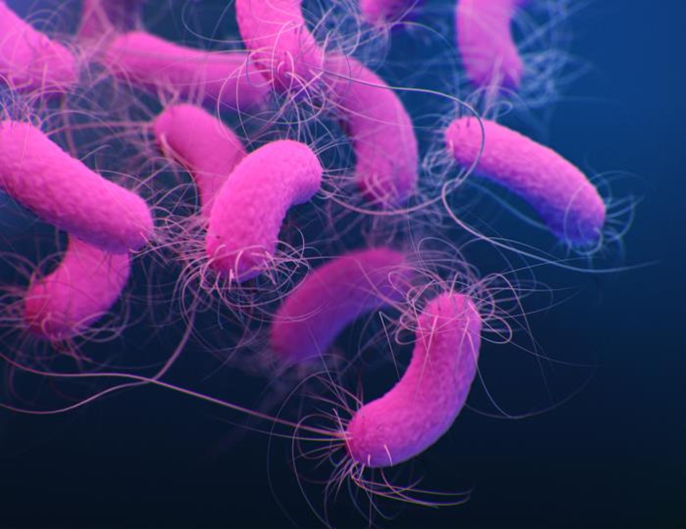
Pseudomonas aeruginosa can cause infections in a variety of places in the human body, including blood, the urinary tract, and lungs. These infections can cause pneumonia and may be fatal. People with weakened immune systems are particularly at risk for P. aeruginosa infections, and those who are on mechanical ventilation, are also highly susceptible. The World Health Organization has named P. aeruginosa as one of the twelve most dangerous bacterial pathogens in the world, highlighting the serious threat it poses to human health.
This study has shown that P. aeruginosa uses several strategies to invade the body; the pathogen can enter the top layers of lung tissue to then attack deeper cells.
Our airways and lungs are lined with a layer of cells that are tightly packed together, and lubricated with mucus that can trap particles and microbes. Special cells remove this mucus, helping to protect us from pathogens. But this barrier is not always impenetrable. Pseudomonas are able to cross it.
With stem cells that are genetically reprogrammed to behave as specific cell types, the researchers created the lung organoids and mimicked the infection process.
"These lung models enabled us to uncover the pathogen's infection strategy. It uses the mucus-producing goblet cells as Trojan horses to invade and cross the barrier tissue. By targeting the goblet cells, which make up only a small part of the lung mucosa, the bacteria can breach the defense line and open the gate," explained study leader Professor Urs Jenal of the Biozentrum at the University of Basel.
The pathogen uses secretion systems to attack and invade goblet cells. Once inside, the bacterial invader replicates over and over, until the goblet cell bursts. This rupture not only releases many more bacterial pathogens that can attack other cells, it also disrupts the tissue barrier, weakening the lung's protection. The pathogenic bacteria then move to this weak spot and target other cells in the area to make the breach larger, so they can infect deeper tissues.
While we now know far more about the infection process, the adaptive behavior of Pseudomonas is still a bit of a mystery. These microbes have to be able to migrate, then launch an attack on goblet cells, then activate virulence factors.
However, the researchers have a way to solve that mystery. They created a biosensor that can track a signaling molecule called c-di-GMP that is used by bacteria to modify cellular activity. This work was reported in Nature Communications.
"This is a technological breakthrough," said Jenal. The investigators will now be able to observe the signaling molecule in real time to determine how it regulates the pathogen, and study lung infections even more closely.
Experienced research scientist and technical expert with authorships on over 30 peer-reviewed publications, traveler to over 70 countries, published photographer and internationally-exhibited painter, volunteer trained in disaster-response, CPR and DV counseling.