Modern bioassays tend to fall under three common modalities: colorimetric, luminometric or fluorometric. Colorimetric assays record the amount of light absorbed by a reporter compound. This absorbance is often presented as optical density (OD). On the other hand, luminometric assays employ luminescent reporter compounds, which produce light. A popular example of this is the luciferase / D-luciferin system. Units for these types of assays are relative light units (RLU). Finally, fluorometric assays utilize reporters which emit light, upon illumination (i.e. excitation) by a secondary light source. This emission is captured in relative fluorescence units (RFU).
The principle of fluorescence is that a substance absorbs energy, in the form of photons, thereby entering an excited state, and as it returns to its ground state, releases that energy back as emitted light. From this, several key aspects can be derived. First, substances have a distribution of wavelengths over which energy is absorbed. This is the substance’s excitation spectrum, the greatest point being its excitation maximum, and the degree to which it absorbs energy being its extinction coefficient. Second, the photon released by a fluorescing substance contains less energy than is absorbed, resulting in an emission light of longer wavelength. This difference between the excitation peak and emission peak is known as the Stokes’ shift, and the efficiency with which a substance converts absorbed energy into emitted energy is known as its quantum yield.
One of the first applications of fluorescence in bioassays was green fluorescent protein (GFP), with an excitation / emission peak of 475 nm / 510 nm, and extinction coefficient / quantum yield of 7000 M-1 cm-1 / 0.79. With over a million publications, GFP is historically significant for the proliferation of 488 nm excitation as a standard instrument light source, “green” as the most commonly captured emission signal, and to date, it is still popular as a reporter gene for cellular studies. Launching from the success of GFP, significant developments have arisen in the field of fluorescence detection, most notably being the increase in availability of synthetic fluorescent compounds, known as fluorophores, which has enabled bioassays ranging from cell viability to PCR.
Cell viability
One of the most common assessments needed in biological research is the determination of cell viability, that is, the degree to which a population of cells are live or dead. To this end, colorimetric assays have been well established. For instance, trypan blue exclusion assays have been used to count dead cell populations. This relies on the principle that trypan blue is cell impermeable, therefore only able to stain dead cells which have compromised cellular membranes. The drawback, however, is that live cells remain unstained, or clear, creating an environment that makes them difficult to assess. A potential remedy is to utilize an assay which quantifies live cell populations. Again, there are established colorimetric assays which perform this function, such as MTT assays or CCK-8 assays. However, these tests create the reverse challenge, where only viable cells are counted while nonviable cells are not. So while colorimetric assays are well established, they have inherent limitations. On the other hand, fluorometric technologies offer a unique solution for this application, by offering bioassays which simultaneously quantitates viable and nonviable cells.
Live dead assays rely on two fluorometric reagents, for instance, calcein am and propidium iodide, to quantitate cell viability. Propidium iodide, similar to trypan blue, is membrane impermeable and excluded from live cells, therefore acting as a reporter of nonviability. Then, to detect live cells, calcein am is used. The am ester functional group on calcein increases its hydrophobicity, allowing it to pass through cell membranes, the degree to which can be further enhanced by detergents such as Pluronic F-127. Importantly, passing through the cell membrane is not in itself an indication of viability, given that calcein am will also permeate cells with compromised membranes. Here, it is worth noting that the am ester group serves another purpose, which is to render the calcein molecule non-fluorescent. Only in live cells, where esterases are ubiquitous and active, is the am ester group cleaved, thereby allowing calcein to fluoresce. In this way, calcein am detects specifically for live cells, and in conjunction with propidium iodide, enables live dead assays to simultaneously assess viable and nonviable cells within the same population, thereby exemplifying one of the key advantages of fluorometric assays, which is allowing multiplex analysis.
Cell imaging
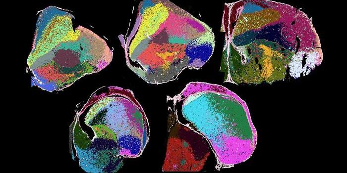
One area where multiplex analysis is particularly important is cell imaging, where the ability to simultaneously visualize different cellular components provides key insights into subcellular interactions and processes. Of priority amongst these compartments is the nucleus, of which a multitude of fluorophores have been developed, for instance, acridine orange and 7-AAD. Acridine orange is a membrane permeable dye that binds to DNA and RNA via intercalation, while 7-AAD binds nucleic acids with a similar principle but is cell impermeable. The benefit of using either probe is that their excitation / emission spectra resemble that of GFP, creating compatibility with preexisting, and prevalent, fluorescence microscopes and flow cytometers. If multiplexing is required, for example, if the green fluorescence channel is already committed to GFP detection, blue fluorescence DNA probes are also commonly available. Amongst these, DAPI and Hoechst 33342 are two popular fluorophores.
In addition to fluorescence imaging of the nucleus, reagents have been developed to label all parts of the cell. For instance, phalloidin conjugated to a fluorophore can be used to visualize actin filaments, which comprise the cytoskeleton. Stains have also been developed for lysosomes and mitochondria. For the latter, because of its importance in cell metabolism and ATP generation, fluorescence assays, such as JC-1, have even been developed to assess mitochondrial membrane potential. For labeling membranes more generally, a family of lipophilic fluorescent stains have found success, namely, DiD, DiI and DiR dyes.
While the aforementioned applications of fluorescence technology focus on structural labeling of cells, there are also a great number of fluorophores which enable functional bioassays. Critical amongst these are the family of calcium indicators, such as Fluo-4 AM and Fluo-8 AM. These allow researchers to study calcium response in cells, which is vital to GPCR-based drug screening and development for the treatment of diseases. In the same vein, fluorophores like thioflavin T have been used in disease research to study the buildup of amyloid fibrils that are a key marker in Alzheimer’s disease.
Macromolecule detection
As illustrated, fluorescence bioassays are powerful tools for cell-based research. Part of this stems from the wide ranging selection of available fluorophores and the diverse number of targets to which they can be conjugated, or chemically labeled. For instance, labeling the annexin v protein with fluorescein yields Annexin V FITC, a reagent useful for monitoring cellular apoptosis through binding of translocated surface phosphatidylserine. Relatedly, conjugating CD antibodies with phycobiliproteins, such as phycoerythrin and allophycocyanin, have proven greatly useful as a means of sorting cells by surface markers.
Another class of important biomolecules that can be conjugated with fluorophores are oligonucleotides, which represent short nucleic acid sequences. These, for instance, can be labeled with Cy3 or Cy5 for use in fluorescence in situ hybridization (FISH), to perform spatial detection of sequence specific nucleic acids within cells. As phosphoramidites, 5-FAM and 6-FAM can easily be incorporated into the synthesis of oligonucleotides which, when paired with an appropriate quencher, can act as a molecular beacon, serving as the basis for TaqMan probes in real time PCR analysis. In workflows that utilize nucleic acid purification techniques, such as capturing mRNA with oligo(dT) magnetic beads, fluorescence can be used as a tool to quantify final yield and assess extraction efficiency.
Although there is already a significant toolkit of fluorometric assays available to researchers, novel methodologies are continually being developed and improved. For instance, brdU has long been used to track cell proliferation by monitoring the incorporation of the dNTP during DNA synthesis. However, the detection has been largely performed through the use of antibodies. New XdU assays bypass the reliance on antibodies, labeling dNTPs directly for fluorescence quantification. Other long standing assays include protein quantification tests, such as the BCA assay and the Bradford assay, which have also seen improvements through replacement by fluorometric methods. As the chemistry behind fluorescence and synthetic fluorophores continues to mature, it is without doubt that more assays will become available, enabling greater and more insightful research into the most pressing biological questions.