The same gene in the brain that codes for a biochemical “self-destruct trigger” in some tissues protects neurons from West Nile virus without causing cell death. From the University of Washington, researchers are studying how the gene, RIPK3, works to keep the peace.

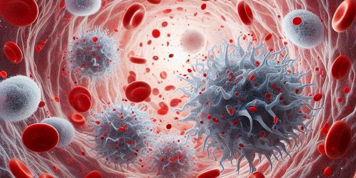
West Nile virus is a mosquito-transmitted disease first detected in the United States in 1999. Infection only develops in about 20-30 percent of people exposed to West Nile virus, and for those who are infected, severe symptoms are very rare. When they do occur though, they include coma, tremors, seizures, and paralysis.
Recent studies in mice show that despite RIPK3’s reputation in other organs of the body, neuron sacrifice is not necessary to eliminate West Nile in the brain and order up a response from the immune system. Instead, the chemical pathway simultaneously preserves neurons and recruits immune chemicals and virus-fighting cells to the scene of the crime.
Instead of sacrificing neurons, RIPK3 activation in the brain results in chemokine recruitment, immune chemicals that in turn attract white blood cells to do the fighting against West Nile virus. These white blood cells eliminate West Nile from the brain not by suppressing viral reproduction, but by causing inflammation. Because of this approach, RIPK3 is likely involved in neurodegenerative disorders and autoimmune disease in the brain. However, targeting RIPK3 as a treatment for these conditions would have to include a careful balance, to ensure the brain is not left unprotected against viruses like West Nile.
"RIPK3 acts as part of the milieu of signals that support anti-viral inflammation in the brain," explained the lead author of the study, Brian Daniels.
During their studies with RIPK3 in mice, researchers procured RIPK3 deficient mice and saw that these mutants were highly susceptible to West Nile virus infection, a direct result of lacking “chemokine-generated neuroinflammation.”
In addition to likely playing a part in autoimmune disease, RIPK3’s unique role in the brain indicates that there may be other central nervous functions for the gene that scientists have yet to discover. Senior author Andrew Oberst says that there’s something special about neurons that must drive RIPK3 to suppress its self-destructive nature in the brain, “perhaps because they are non-renewable and too important to undergo cell death.”
The present study was published in the journal Cell.
Sources: CDC, European Centre for Disease Prevention and Control, University of Washington Health Sciences